Here you can find an overview of a few of the research projects I have worked on between 2019-2021. For further information or if you have any questions, please do not hesitate to contact me.
Collaborations with visitors of the Advanced Imaging Center
 Between 2019 and 2021, I have supported (inter)national scientists and Janelians at HHMI Janelia Research Campus with their imaging experiments at instruments at Janelia’s Advanced Imaging Center (AIC), Janelia’s Light Microscopy Core, and other instruments on campus. The support reached from technical consultations focusing on experimental design, over support, and guidance during imaging experiments at the respective microscopes to support with the processing and analysis of the acquired data during and after the imaging sessions. Projects at the AIC were particularly demanding since the AIC instruments are solely pre-commercial systems (iPALM, Lattice Light Sheet Microscope, SiMView Light Sheet Microscope, Aberration Corrected Multifocus Microscope, MOSAIC, etc.) that require more hands-on support and troubleshooting. In total, I have helped over 15 individual imaging projects at the AIC, ranging from imaging experiments of algae at Janelia’s iPALM with nanoscale resolution to imaging experiments of the whole living zebrafish at Janelia’s LLSM and SiMView. The diversity of different projects at various, particularly pre-commercial, instruments requires expertise in molecular biology, optical engineering, hardware design, programming, data analysis, and data visualization, which perfectly suits with my previous training.
My AIC work with visitors resulted in the following papers:
- Nogueira, A.T., Herron J.C., O’Shaughnessy E.C., Boehm U., et al.: “Resolving protein conformation in iPALM.” Biophysical Journal (2023), submitted
- Reiche, M.A., Aaron J., Boehm U., et al.: “When Light Meets Biology: How the Specimen Affects Quantitative Microscopy. “ J. Cell Sci. (2022), DOI:10.1242/jcs.259656
- Boehm U. “Janelia+EMBL BioImaging Seminar Series: How We Started a Successful Seminar Series during the Pandemic” Focalplane (2022), DOI:https:10.1242/focalplane.6011
- Boehm U., Galbraith C.: “Extending the performance capabilities of isoSTED” Biophysical Journal 120 (2021), p1-3, DOI:10.1016/j.bpj.2021.07.005
- Galbraith J., Aaron J., Boehm U., Chew T.-L. and Galbraith C.: “Resolving the 3D Nano-architecture of the Actin Cytoskeleton” Microscopy and Microanalysis (2020), p1, DOI:10.1017/S1431927620016736
Integrating machine learning strategies for iPALM
 Single-molecule localization microscopy (SMLM) has had remarkable success in imaging cellular structures with nanometer resolution, but standard analysis algorithms require sparse emitters, which limits imaging speed and labeling density. In 2020, I had the pleasure to support the development and distribution of DECODE. This computational tool can localize single emitters at high density in three dimensions with highest accuracy for a large range of imaging modalities and conditions to overcome this limitation using deep learning. To support the adoption of DECODE, we created DECODE tutorials for the virtual I2K workshop at the Janelia Research Campus.
This project resulted in the following paper:
- Speiser A., Müller L.-R., Hoess P., Matti U., Obara C.J., Legant W.R., Kreshuk A., Macke J.H., Ries J., Turaga S.C. (2021): “Deep learning enables fast and dense single-molecule localization with high accuracy” Nature Methods, 44, p7508–7510, DOI:10.1038/s41592-021-01305-1
Snapshop Hyperspectral MFM
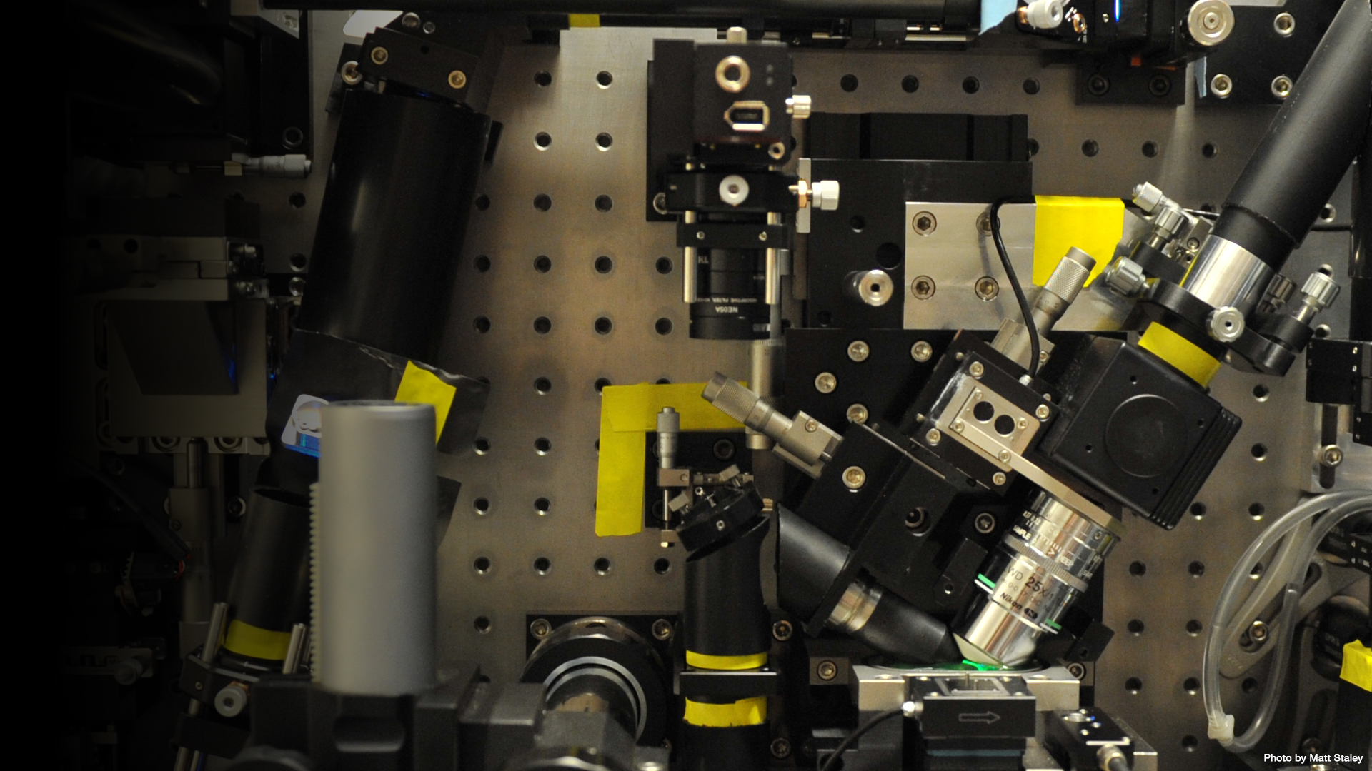
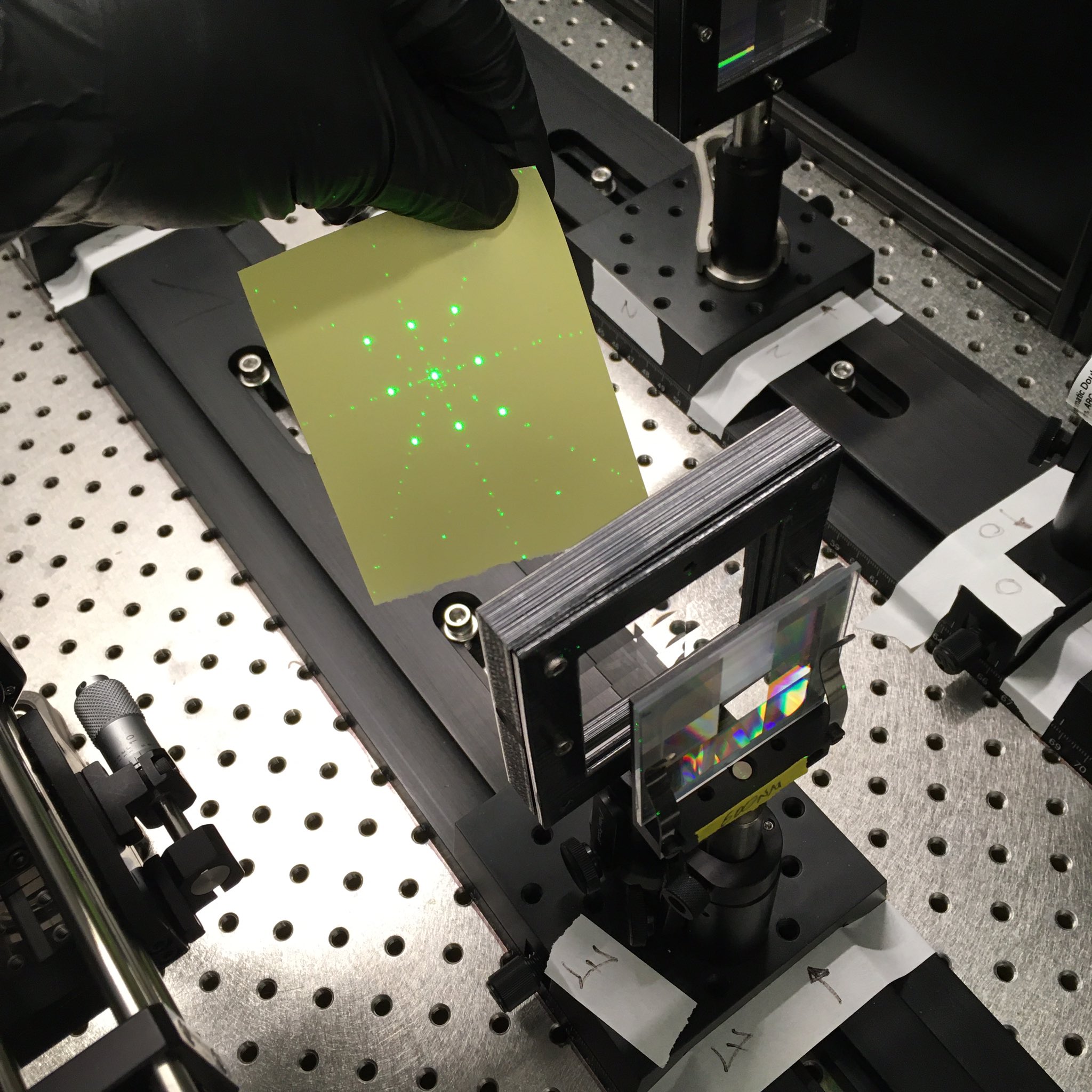 After decommissioning the AIC’s Multifocus Microscope (MFM), I repurposed the system. Originally, it was able to quickly capture multiple z planes at once on different segments on a single camera by optical manipulation (i.e., a novel diffraction grating design and chromatic correction scheme are appended to the camera port of a high-resolution epifluorescence microscope). I converted it into a spectral instrument that allowed capturing multiple spectral components of a single z plane at once on different segments on a single camera. Therefore, new optical elements needed to be designed, built, and integrated into the old system. Along the way, new alignment tools were also developed. The project is still ongoing.
This project resulted in the design of several new alignment tools for optical system. The design of a laser alignment tool was published as Microscope Breadboard Laser Alignment Tool on Janelia’s Open Hardware portal and later on incorporated as Breadboard Mountable Beam Height Ruler in Thorlabs portfolio:
- Boehm U. (2021): “Microscope Breadboard Laser Alignment Tool” Janelia Open Hardware portal
- Boehm U. (2022): “Breadboard Mountable Beam Height Ruler” Thorlabs, Inc.
Reporting and reproducibility in microscopy
 Microscopes have long been a workhorse in the life sciences. Despite their abundance and how familiar scientists are with seeing microscopy data, there are still significant gaps in how microscopy experiments are reported. These gaps limit how readers of published work can assess the data and can hobble reuse and reproducibility.
During the pandemic in 2020 and 2021, I participated in multiple microscopy initiatives (QUAREP-LiMi, BioImaging North America, etc.) to work on guidelines and tools for improving the tracking and reporting of microscopy metadata with an emphasis on reproducibility and data reuse.
This project resulted in the following papers:
- Gaudreault N., …, Boehm U., et al.: “Illumination Power and Illumination Stability” protocol.io (2022), DOI:10.17504/protocols.io.bzp8p5rw
- Rigano A., …, Boehm U., et al.: “Micro-Meta App: an interactive tool for collecting microscopy metadata based on community specifications” Nature Methods (2021), DOI:10.1038/s41592-021-01315-z
- Hammer M., Huisman M., Rigano A., Boehm U., et al.: “Towards community-driven metadata standards for light microscopy: tiered specifications extending the OME model” Nature Methods (2021), DOI:10.1038/s41592-021-01327-9
- Boehm U.*, Nelson G.*, et al.: “QUAREP-LiMi: A community-driven initiative to establish guidelines for quality assessment and reproducibility for instruments and images in light microscopy” Journal of Microscopy (2021), DOI:10.1111/jmi.13041
- Boehm U.*, Nelson G.*, et al.: “QUAREP-LiMi: a community endeavor to advance quality assessment and reproducibility in light microscopy” Nature Methods (2021), DOI:https://doi.org/10.1038/s41592-021-01162-y
For further information or if you have any questions, please do not hesitate to contact me.