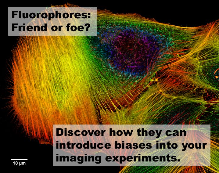
Fluorescence microscopy is a powerful tool that allows scientists to visualize & study biological structures with incredible detail. However, the fluorophores we use to illuminate our target structures can introduce several biases that may lead to misinterpretation of data. Ultimately, we are visualizing our target structures indirectly through fluorophores, and it is essential to take this into account.
Throughout the past 15 years, I have not only built plenty of microscopes but also supported hundreds of scientists with their imaging experiments, and I would like to emphasize a few of these common biases that often require careful consideration:
Photobleaching
Photobleaching occurs when fluorophores lose their ability to fluoresce after prolonged exposure to light. This can lead to a decrease in signal intensity over time, potentially causing us to underestimate the presence or abundance of certain molecules.
Linker Length and Composition
The length & composition of the linker used to attach the fluorophore to the targeting molecule can affect the proximity of binding. Shorter linkers generally result in closer binding, while longer linkers may introduce spatial separation, leading to errors in spatial resolution & potentially misrepresenting the biological architecture.
Label Density
The density of fluorophore labels can significantly impact our observations. Low label density might result in incomplete visualization, missing out on key details.
Polarization Effects
Fluorophores can exhibit polarization-dependent behavior, meaning their emission intensity can vary based on the orientation of the light source & the fluorophore itself. This can introduce variability in the data, especially when comparing different samples or experimental conditions.
Spectral Overlap
Different fluorophores can have overlapping emission spectra, leading to cross-talk between channels. This can complicate the interpretation of multi-color experiments, where distinguishing between signals from different fluorophores becomes challenging.
Environmental Factors
The local environment around fluorophores, such as pH, temperature, & the presence of quenching agents, can affect their fluorescence properties. These factors can introduce variability & potentially skew the data.
Conclusion
As imaging scientists, it’s crucial to be aware of these biases & take them into account when designing your imaging experiments & analyzing your data. By understanding the limitations & potential pitfalls of fluorescence microscopy, you can make more accurate & reliable interpretations of your imaging experiments. Wishing you the best of luck with your imaging experiments!
Feature photo: Depth Coded Phalloidin Stained Actin Filaments Cancer Cell, CC BY-SA 3.0, via Wikimedia Commons
For further information or if you have any questions, please do not hesitate to contact me.


